前面我们在推文 细胞亚群的特异性标记基因也许真的很难提到的Cancer-associated fibroblasts (CAFs)是比较难以精确的细分亚群。而且我们讨论了[T细胞可以简单分成4类](https://mp.weixin.qq.com/s/A_lIKqboCDUSpbyWPSGf-Q),其实CAFs的分类也值得深入讨论!
首先,在小鼠里面可以看到,myofibroblast (myCAF), inflammatory fibroblast (iCAF), and antigen-presenting fibroblast (apCAF),但是在人类里面并没有很明显的apCAF,也就是说通常是分2类,很有意思的,值得分享!比如,于AUGUST 2019发表在 CANCER DISCOVERY 杂志的文章:《Cross-Species Single-Cell Analysis of Pancreatic Ductal Adenocarcinoma Reveals Antigen-Presenting Cancer-Associated Fibroblasts》,链接是:https://cancerdiscovery.aacrjournals.org/content/9/8/1102.long
最后得到了两个CAFs亚群,分别是:
- CAFs expressing high levels of αSMA, which we therefore named “myofibroblastic CAFs” (myCAF),
- CAFs expressing low levels of αSMA but high levels of cytokines and chemokines, which we named “inflammatory CAFs” (iCAF;)
而且全文就是重点讨论了CAFs的两个亚群的功能, 就是myofibroblast (myCAF) 和 inflammatory fibroblast (iCAF)。这个课题的单细胞转录组数据共有2万多个细胞,The human RNA-seq data are available at NCBI dbGaP under the accession number phs001840.v1.p1. 不方便下载。样品来源于:Six tumors from patients with untreated PDAC and adjacent-normal pancreas tissues from two of these patients,分成15个细胞亚群,其中962 fibroblasts仍然是可以分成两个亚群,都表达COL1A1, FAP等CAFs的标志基因,但是这两群又有所差异,如下所示:
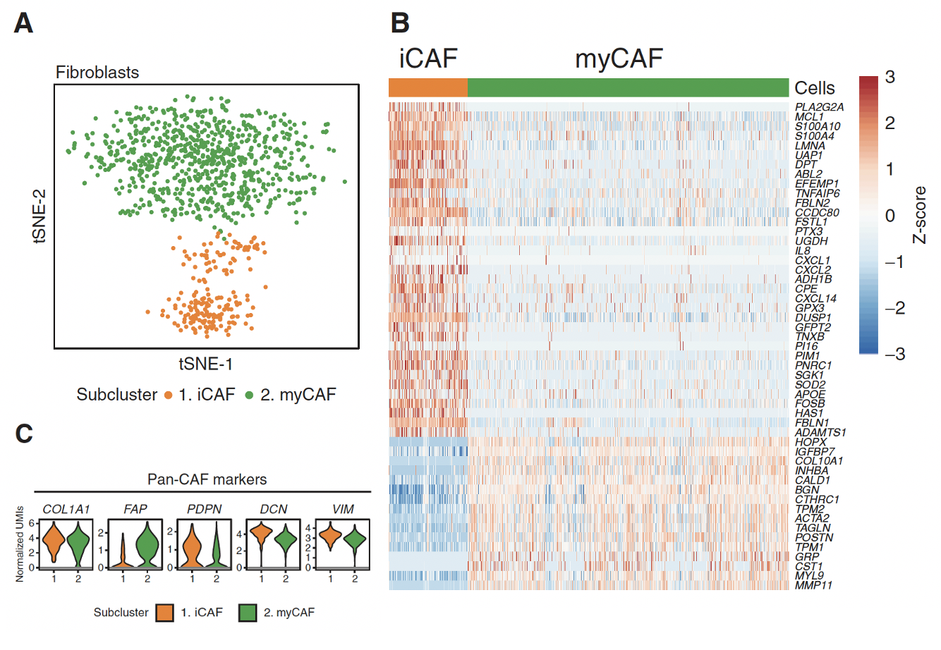
两个亚群各自的标志基因是:
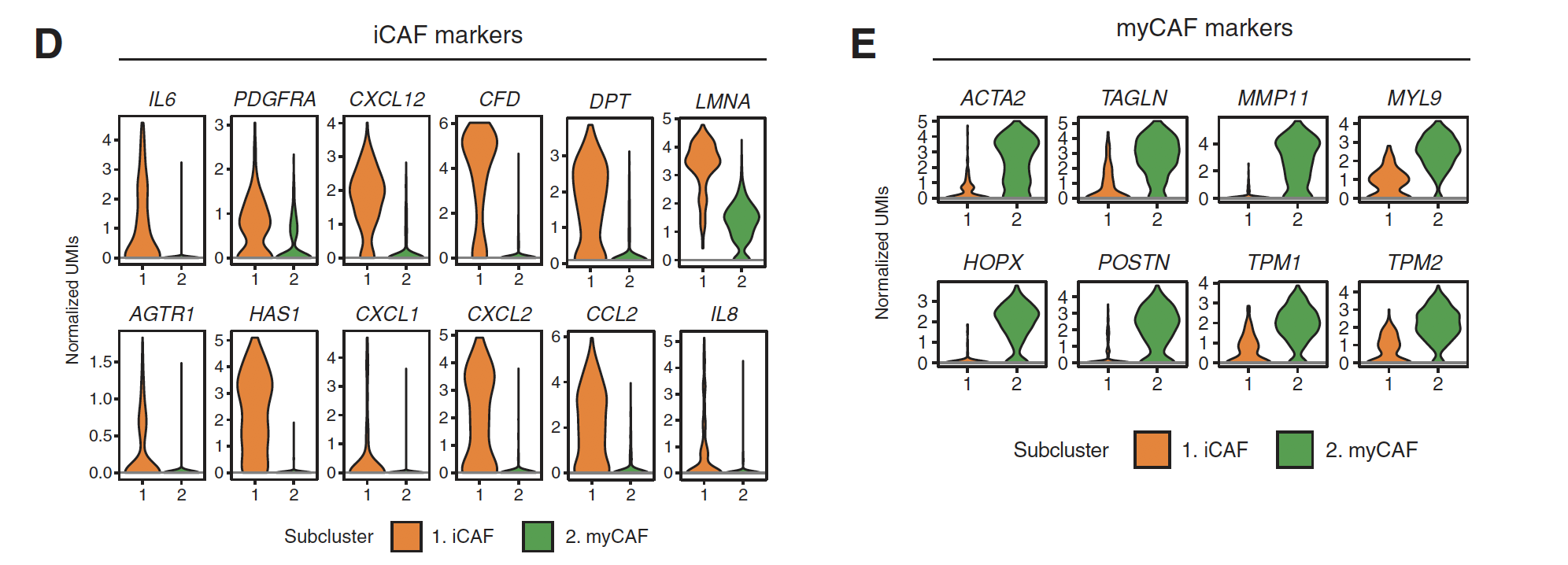
差异还是很明显的!
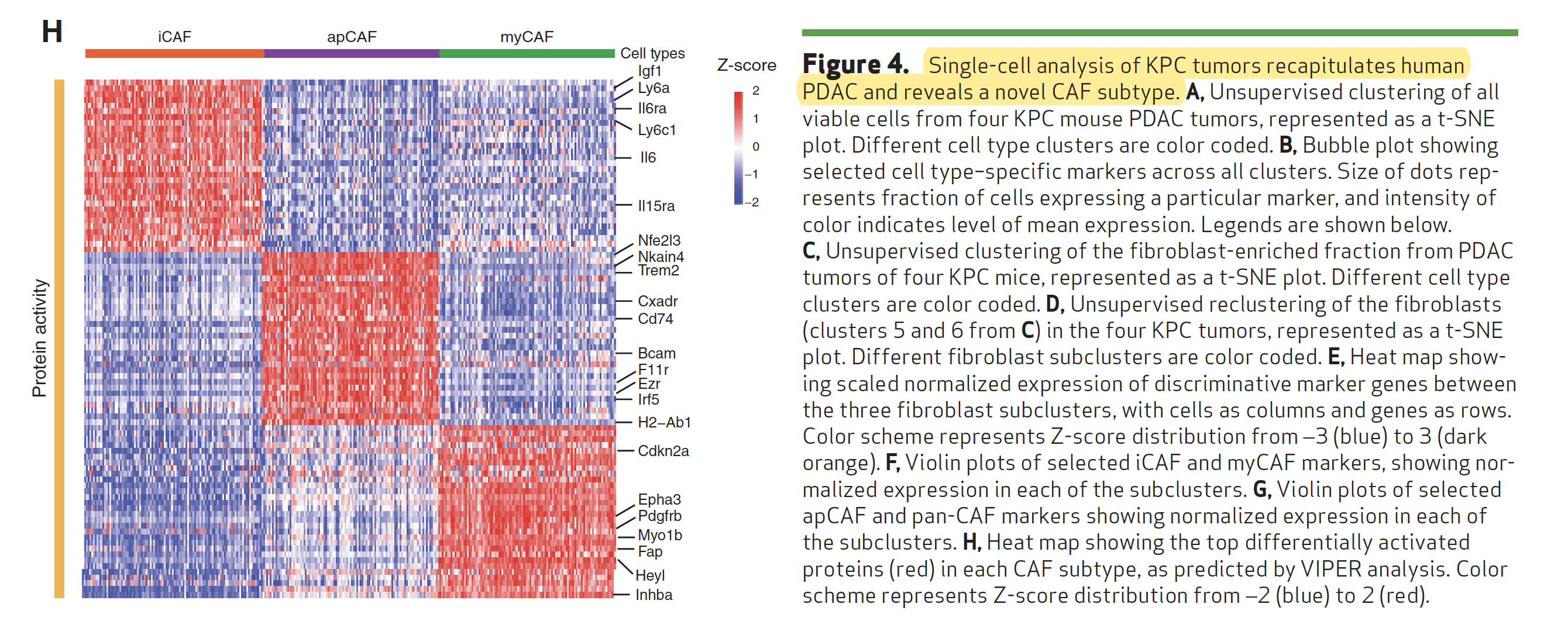
在肿瘤小鼠模型的单细胞数据里面可以分3群
肿瘤小鼠模型数据是:We combined the sequencing data from the fibroblast-enriched populations isolated from the four KPC tumors into one data set consisting of 8,443 cells and analyzed them together.
数据在:https://www.ncbi.nlm.nih.gov/geo/query/acc.cgi?acc=GSE129455
GSM3713177 KPC1_Viable
GSM3713178 KPC2_Viable
GSM3713179 KPC1_FibroblastEnriched
GSM3713180 KPC2_FibroblastEnriched
GSM3713181 KPC3_Viable
GSM3713182 KPC4_Viable
GSM3713183 KPC3_FibroblastEnriched
GSM3713184 KPC4_FibroblastEnriched
多了的这个Cancer-associated fibroblasts (CAFs)亚群命名为:“antigen-presenting CAFs” (apCAF).
使用ARACNe and VIPER algorithms.,清晰可见:
另外一种可视化方法如下:
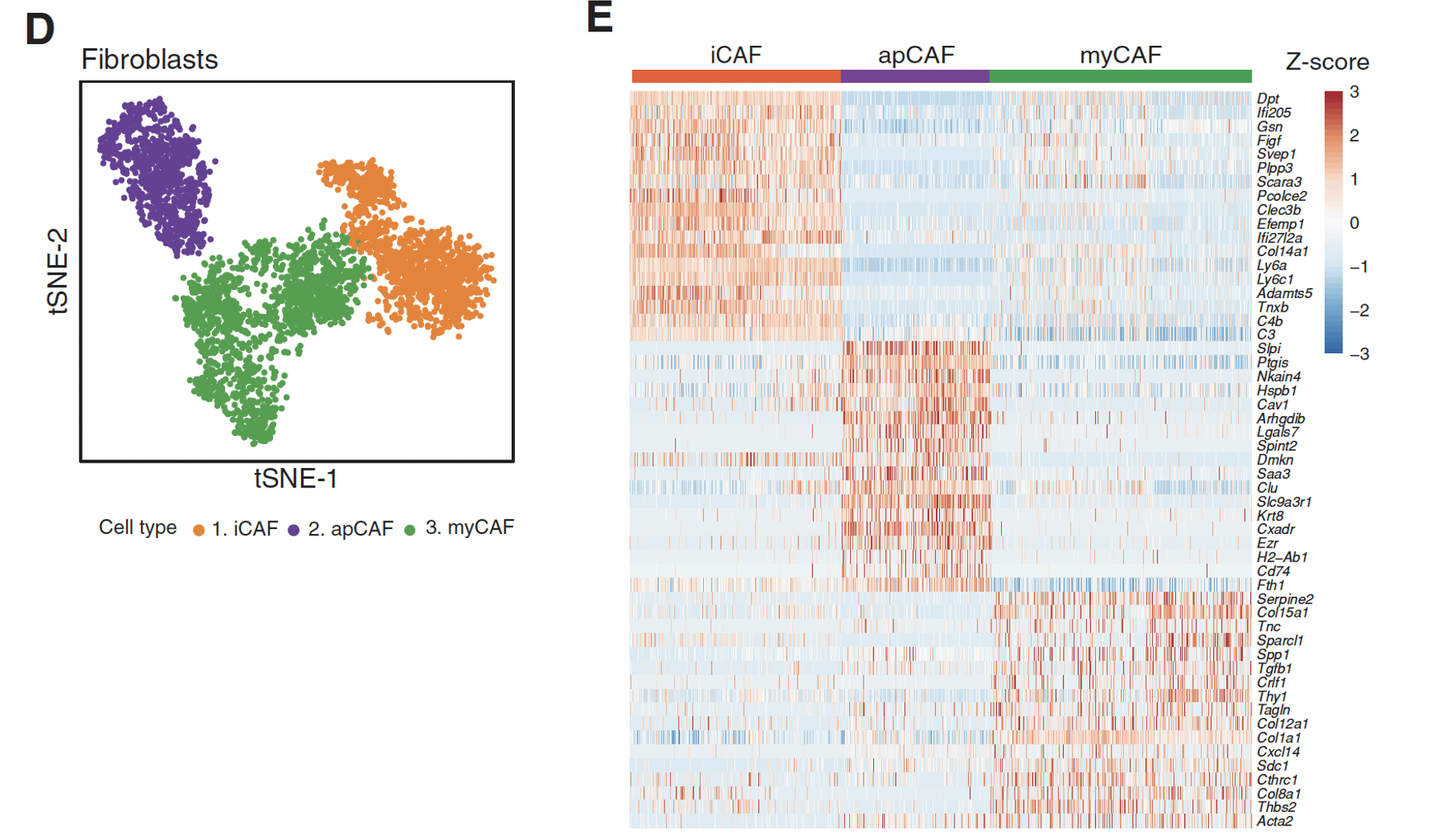
确实是各自的marker基因很清晰,“antigen-presenting CAFs” (apCAF) 就特异性表达 :
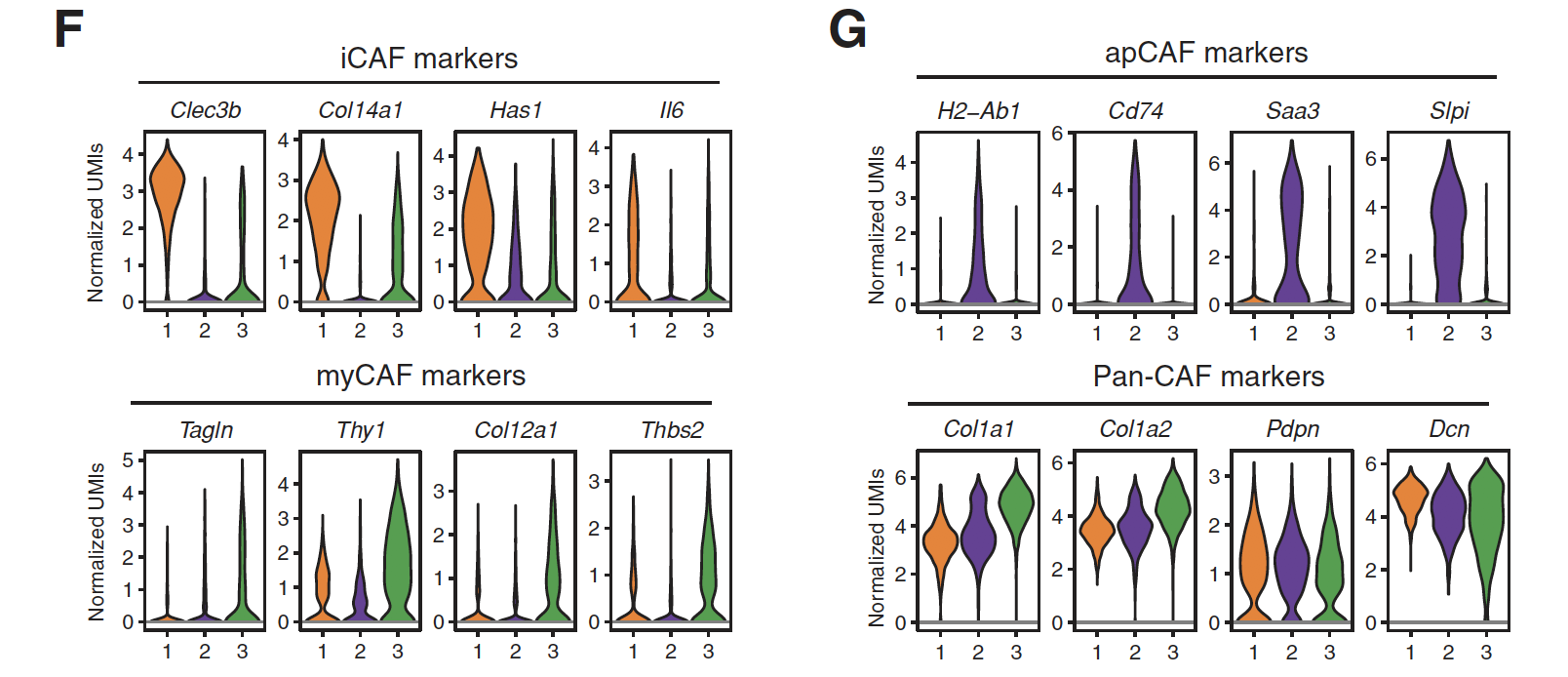
GSEA分析发现这3群也有各自的通路激活情况:
- upregulation of inflammatory pathways in mouse iCAFs (e.g., JAK/STAT signaling, cytokine interactions with their receptors and coagulation;
- The pathways specifically upregulated in mouse myCAFs were EMT, myogenesis, ECM receptor interaction, and focal adhesion
- 而apCAF subtype,独特的通路包括:antigen presentation and processing, fatty-acid metabolism, MYC targets, and MTORC1 signaling
但是为什么前面在人类单细胞数据里面没有发现apCAF呢
前面提到962 fibroblasts仍然是可以分成两个亚群,都表达COL1A1, FAP等CAFs的标志基因,那我们检查看看apCAF subtype的标志基因,如下:
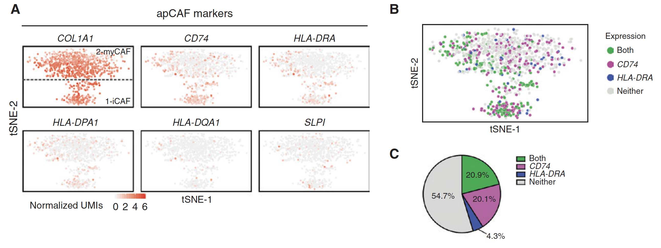
只能说姑且认为是两个物种的差异?或者说小鼠模型本来就不可能是一模一样的模拟人类的肿瘤微环境。
看看Cancer-associated fibroblasts (CAFs)在原位肿瘤和转移瘤的差异情况
2020年9月的文章:《Single-cell transcriptome analysis of tumor and stromal compartments of pancreatic ductal adenocarcinoma primary tumors and metastatic lesions》,链接是:https://genomemedicine.biomedcentral.com/articles/10.1186/s13073-020-00776-9
总共是 16个10x单细胞数据:https://www.ncbi.nlm.nih.gov/geo/query/acc.cgi?acc=GSE154778
GSM4679532 P01: Primary tumor 01 (10x 3' v1)
GSM4679533 P02: Primary tumor 02 (10x 3' v2)
GSM4679534 P03: Primary tumor 03 (10x 3' v2)
GSM4679535 P04: Primary tumor 04 (10x 3' v2)
GSM4679536 P05: Primary tumor 05 (10x 3' v2)
GSM4679537 P06: Primary tumor 06 (10x 3' v2)
GSM4679538 P07: Primary tumor 07 (10x 3' v2)
GSM4679539 P08: Primary tumor 08 (10x 3' v2)
GSM4679540 P09: Primary tumor 09 (10x 3' v2)
GSM4679541 P10: Primary tumor 10 (10x 3' v2)
GSM4679542 MET01: Metastatic lesion 01 (10x 3' v2)
GSM4679543 MET02: Metastatic lesion 02 (10x 3' v2)
GSM4679544 MET03: Metastatic lesion 03 (10x 3' v2)
GSM4679545 MET04: Metastatic lesion 04 (10x 3' v2)
GSM4679546 MET05: Metastatic lesion 05 (10x 3' v2)
GSM4679547 MET06: Metastatic lesion 06 (10x 3' v2)
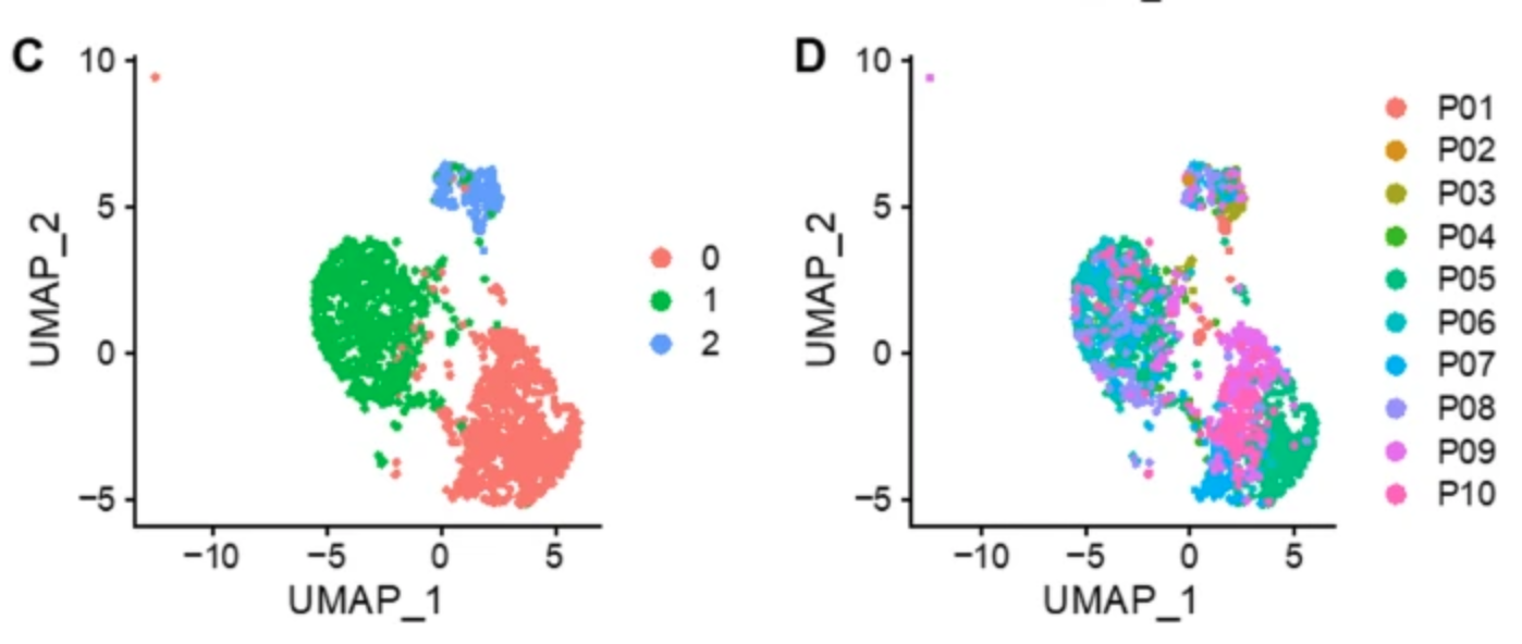
同样的,可以进行基本的降维聚类分群,参考前面的例子:人人都能学会的单细胞聚类分群注释 ,第一次分群就非常漂亮!图例是:
- c Three major clusters are formed by CAFs from primary tumors.
- d CAFs from different patients are mixed in the different clusters
描述是:
Figure 2c shows the unsupervised clustering of 1753 CAF cells identified in Fig. 1a. The CAFs formed 3 major clusters: c0, c1, and c2 (Fig. 2c). Unlike the tumor cells, the CAFs did not cluster by patient.
这篇文章也提到了:Elyada and colleagues previously described 3 subtypes of CAFs identified in PDAC tumors: myofibroblasts (myCAFs), inflammatory fibroblasts (iCAFs), and antigen-presenting fibroblasts (apCAFs)
参考:
- Semin Cancer Biol . 2020 May; 《Cancer-associated fibroblasts in desmoplastic tumors: emerging role of integrins 》, https://pubmed.ncbi.nlm.nih.gov/31415910/
- https://cancerdiscovery.aacrjournals.org/content/9/2/173
- https://www.thelancet.com/journals/ebiom/article/PIIS2352-3964(20)30030-X/fulltext
- Am J Physiol Cell Physiol . 2020 Aug 1; 《Recent advances in understanding cancer-associated fibroblasts in pancreatic cancer》 ,https://pubmed.ncbi.nlm.nih.gov/32432930/
- J Exp Med. 2017 Mar 6; 《Distinct populations of inflammatory fibroblasts and myofibroblasts in pancreatic cancer》
- https://onlinelibrary.wiley.com/doi/epdf/10.1111/cas.14537
- GSE125588
- GSE129455
- Yin & Yang: The Duality of Cancer-Associated Fibroblasts in Pancreatic Cancer
- Stromal cells affect immune infiltrate in triple-negative breast cancer